Technique :
The setting of the sequence's parameters is a compromise meanwhile of acquisition and resolution. One obtains as the case may be a "T1", "T2" or "gradient echo" sequence.
Each sequence requires 4 to 8 minutes of acquisition depending on the number of slices and the resolution.
The contrast media is Gadolinium, a rare earth with exceptional side effects.
Fat suppression techniques (Fat-Sat, STIR, SPIR,...) improve the contrast after injection of Gadolinium (figure 32b).
Angiographic sequences by MR (MRA) are realized with (better resolution) or without injection of Gadolinium, and are based on fast 2D or 3D "gradient echo" acquisition protocols (in one breathhold).
Results :
The semiology is variable depending on the T1 , T2 or EG weight of each sequence; each structure having its own curves of signal decreasing after a RF excitement :
- The fat appears in iso or hyper signal on T1 or T2 sequences.
- Vessels with slow stream are in T1 hypersignal.
- Vessels with fast stream are in T1 hyposignal.
- After injection of gadolinium, the arterial or venous vessels are in hypersignal on T1 weighted sequences.
- Lymph nodes appear in T1 hyposignal
Figure 32: Abdominal IRM. Axial native cups(cuttings). A) images balanced in T1. The fat appears in hypersignal; the structures vascular and ganglionic in hyposignal ( no injection) b) images balanced in T1 after Gadolinium's injection ( vascular raising) and abolition(deletion) of the signal of the fat (the fat appears in hyposignal, what improves the vascular contrast)
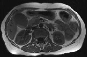
Figure 32 : Abdominal MRI. Native
axial slices.
a) T1. Fat is hypersignal ; vessels and lymph nodes in hyposignal.(no injection)
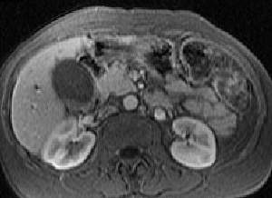
b) Gadolinium enhanced T1 sequence with fat suppression (fat is hyposignal improving the vascular contrast)