One manually unwinds the small bowel and one can observe the fan-shaped aspect of the mesentery, its smooth, regular aspect.
The mesentery is fine and mobile in periphery; more thick and fixed at the proximal edge.
Except with the projection represented by the vessels, one can not individualize the mesentery of the ascending mesocolon.
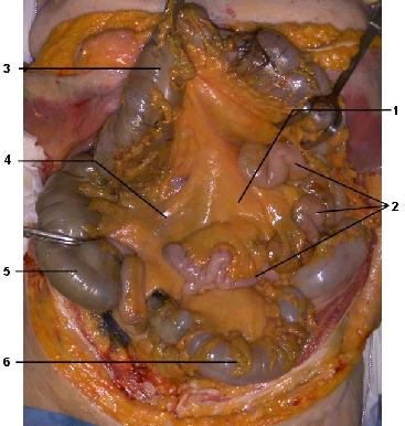
Figure 18 : Anterior view of abdominal cavity. Transverse colon and transverse
mesocolon are reclined up.
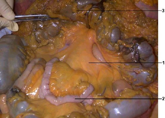
Figure 19 : Antero lateral view (detail of fig.18) of
mesentery
1 Mesentery
2 Small bowel (jéjunum)
3 Transverse colon
4 Ascending mesocolon
5 Right colon
6 Sigmoid colon
In order to individualize the mesentery for a radiologic study, we ligate the pylorus, the right colon beyond the caecum, and resect the pancreas head with duodenum.
The last stage consists in splitting the ascending mesocolon and mesentery as well as cutting mesenteric vessels. (figure 21)

Figure 20 : Anterior view of an anatomic sample containing mesentery, duodenopancreatic block, the whole small bowel, caecum and appendix.
1 Pylore ligatured
2 Jejunum
3 Mesentery
4 Ileon
5 Last ileal loop
6 Appendix and mesoappendix
7 Caecum ligatured